Kleiner Lab devises new approach to spatially map RNA modifications in cells
The study of RNA has exploded in the past decade with advancements that are chipping away at the crucial role these biomolecules—once considered a mere sidenote to DNA—play in human biology. As with any burgeoning field, there is a long road to complete understanding.
This week the Kleiner Lab introduces a new advancement towards this end with a combination approach that allows scientists to peer deeply into the cellular microenvironment and pinpoint RNA modifications spatially. Such precision has been difficult if not unworkable in the past.
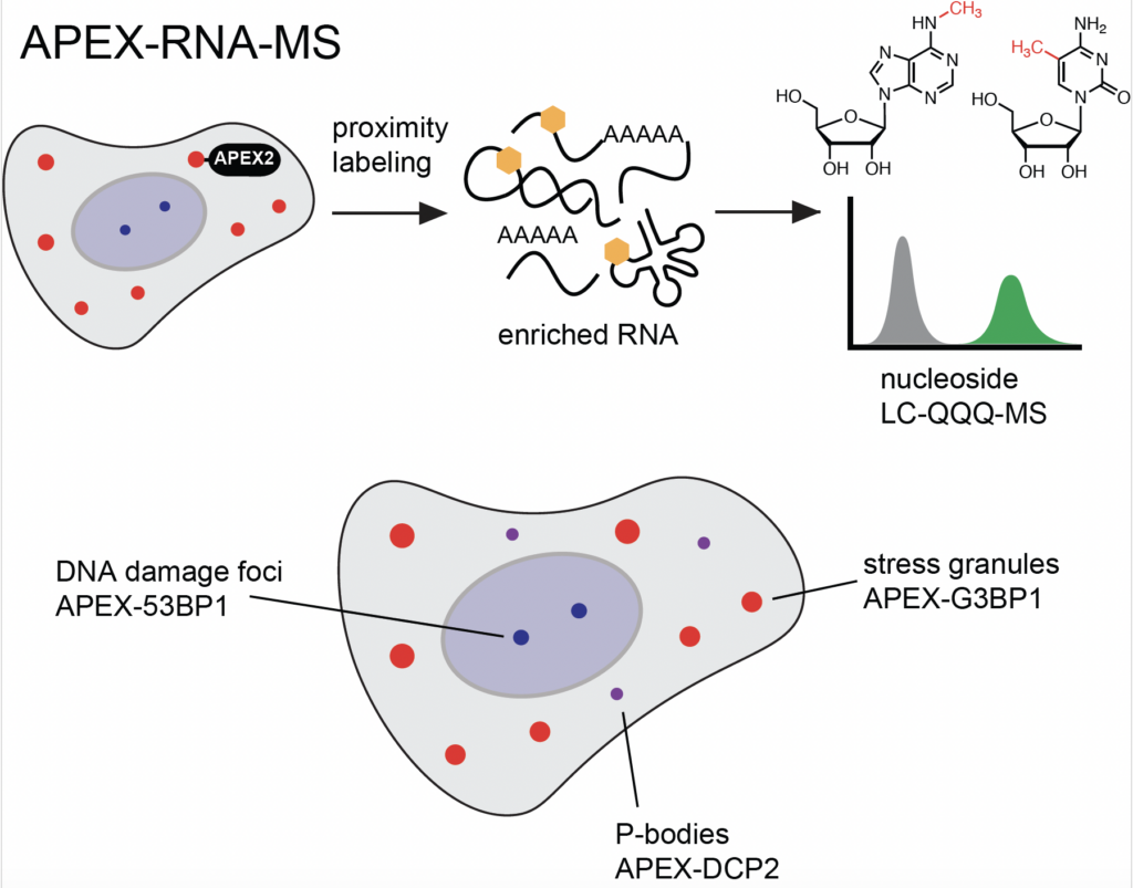
In what is likely the first example of proximity labeling used to identify RNA modifications, researchers married two powerful tools, APEX2 proximity labeling and RNA mass spectrometry, to obtain game-changing subcellular resolution.
Since location and function are intricately linked, the lab’s APEX-RNA-MS approach could illuminate questions surrounding the dynamics of RNA modifications, which feature prominently in gene regulation and cellular processes.
The lab’s research, Quantitative profiling of RNA modifications enriched in non-membrane bound cellular structures using APEX-RNA-MS, appears this week in Cell Chemical Biology.

Graduate student Dhruv Dhingani and author on the Kleiner Lab's latest research, out in Cell Chemical Biology this week.
“We paired proximity labeling with RNA mass spectrometry, in particular ribonucleoside liquid chromatography-mass spectrometry (LC-MS), which has been a workhorse method for RNA modification analysis in our group for many years,” said Associate Professor Ralph Kleiner.
“Our goal was to demonstrate that RNA modifications are involved in the biological processes mediated by biomolecular condensates, which are non-membrane bound structures that concentrate RNA, proteins, and other biomolecules in spatially distinct cellular neighborhoods, ” said Kleiner. “We chose structures with known biological functions, like DNA damage foci, and those with largely unknown functions, like stress granules and P-bodies. We wanted to cast a wide net across diverse biological systems, and also demonstrate the generality of our approach.”
Kleiner noted that he drew inspiration for the research from the Alice Ting Lab at Stanford University, which developed the APEX approach and previously applied it for RNA sequencing, along with proximity labeling-based methods and biological studies emerging from the David MacMillan and the Tom Muir labs at Princeton.
RNA mass spectrometry is an analytical technique that is highly challenging; few labs have the expertise to use it for advancements like this one. It has long been a staple of the Kleiner lab.
“To our knowledge, we are the first to combine proximity labeling with RNA LC-MS to study RNA modifications. RNA LC-MS is a workhorse approach used in various projects in the Kleiner lab. Its high sensitivity and generality make it the perfect tool for looking at RNA modifications that are present at low abundance,” said Dhruv Dhingani, a rising fifth-year graduate student in the lab and an author on the paper.
“This is a very good hypothesis-forming approach, where a lot of the data you get will help lead you to new ideas or to make hypotheses about what RNA modifications are doing in the cell, what their roles are. RNA modifications are still kind of a black box. We know they exist. We know they are different and they change when certain events occur. But we don’t know the purpose for a lot of them. So now we can look at where they are in the cell. It could lead you down the road to why they’re there to begin with.”
A generalized, two-step approach
There are over 150 known RNA modifications, all with diverse chemical structures. Distinguishing these complex “invisible” chemical changes from actual RNA canonical bases and investigating their biological function has been difficult in the past.
The Kleiner Lab approach changes that, making for a clean, two-step technique that is generalizable for other scientists interested in studying RNA modifications and their context-specific roles in cellular biology.

Former Kleiner Lab graduate student Kyung “Josh” Seo *23, first author on the paper.
The lab’s research focused on three non-membrane protein condensates, measuring RNA modifications associated with stress granules, P-bodies, and DNA damage sites to demonstrate that RNA modifications are involved in the biological processes mediated by these condensates.
Essentially, the researchers painted RNAs with a spatially localized APEX enzyme to capture those that occur within the biomolecular condensate. Next, they collected all of the flagged RNAs extant in the cell. Finally, they chopped them into single nucleotides and used nucleotide mass spectrometry to elucidate which modifications are present and quantify their concentration.
In the future, the lab intends to use their approach to study RNA modifications associated with disease-relevant condensates in neurodegeneration and in cancer.
“With our method, we barely scratched the surface of the biology of non-membrane bound granules,” said Kyung “Josh” Seo, who earned his Ph.D. under Kleiner in 2023 and is the first author on this research. “It’s thrilling to know that this approach could reveal much of the mysteries of other cellular structures, as well as those in disease-relevant cell models including neurons and immune cells.”
This research was supported by funding from the National Institutes of Health (R01 GM132189) and the National Science Foundation (MCB-1942565).