Hecht/Scholes Create Room Temp Quantum Dots Using A Novel Protein
Nature uses 20 canonical amino acids as building blocks to make proteins, combining their sequences to create complex molecules that perform biological functions.
But what happens with the sequences not selected by nature? And what possibilities lie in constructing entirely new sequences to make novel, or de novo, proteins bearing little resemblance to anything in nature?
That’s the terrain the Hecht Lab works in. Recently, their curiosity for designing their own sequences paid off in spades.
They discovered the first known de novo protein that catalyzes the synthesis of quantum dots. Quantum dots are fluorescent nanocrystals used in electronic applications from LED screens to solar panels.
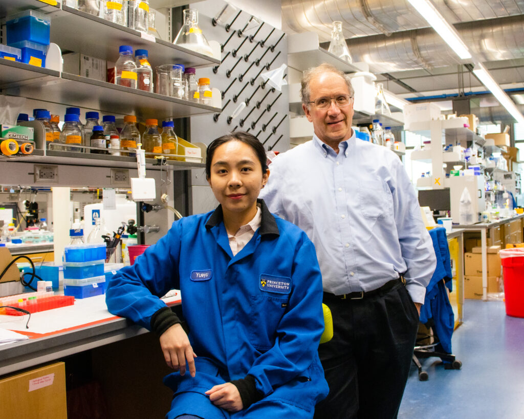
Yueyu Yao, fifth-year graduate student and co-author on the paper, and Professor Michael Hecht in Frick Lab.
The team’s work opens the door to making other nanomaterials in a more sustainable way by demonstrating that protein sequences not derived from nature can be used to synthesize functional materials – with pronounced benefits to the environment.
Quantum dots are normally made in industrial settings using high temperatures and toxic, expensive solvents – a process that is neither economical nor environmentally friendly. Researchers pulled this process off at the bench using water as a solvent, making a stable end-product at room temperature.
“We’re interested in making life molecules, proteins, that did not arise in life,” said Professor of Chemistry Michael Hecht, who led the research with Greg Scholes, the William S. Tod Professor of Chemistry. “In some ways we’re asking, are there alternatives to life as we know it? All life on earth arose from common ancestry. But if we make lifelike molecules that did not arise from common ancestry, can they do cool stuff?
“So here, we’re making novel proteins that never arose in life doing things that don’t exist in life.”
The team’s process can also tune nanoparticle size, which determines the color quantum dots glow, or fluoresce, in. That holds possibilities for tagging molecules within a biological system, for instance staining cancer cells in vivo.
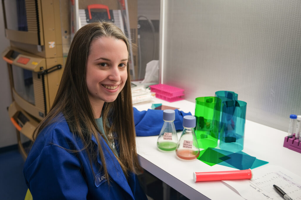
Leah Spangler, lead author on the paper, in Frick Lab last year.
“Quantum dots have very interesting optical properties due to their sizes,” said Yueyu Yao, co-author on the paper and a fifth-year graduate student in the Hecht Lab. “They’re very good at absorbing light and converting it to chemical energy – that makes them useful for being made into solar panels or any sort of photo sensor.
“But on the other hand, they’re also very good at emitting light at a certain desired wavelength, which makes them suitable for making LED screens.”
And because they’re small – comprised of only about 100 atoms and maybe 2 nanometers across – they are able to penetrate some biological barriers, making their utility in medicines and biological imaging especially promising.
The Hecht Lab’s research, “A de novo protein catalyzes the synthesis of semiconductor quantum dots,” was published this week in the Proceedings of the National Academy of Sciences (PNAS).
Why use de novo proteins to make quantum dots?
“I think using de novo proteins opens up a way for designability,” said Leah Spangler, lead author on the research and a former postdoc in the Scholes Lab. “A key word for me is ‘engineering.’ I want to be able to engineer proteins to do something specific, and this is a type of protein you can do that with.
“The quantum dots we’re making aren’t great quality yet, but that can be improved by tuning the synthesis,” she added. “We can achieve better quality by engineering the protein to influence quantum dot formation in different ways.
Based on work done by Sarangan Chari, senior chemist and a corresponding author, the team used a de novo protein it designed named ConK to catalyze, or drive, the reaction. Researchers first isolated ConK in 2016 from a large combinatorial library of proteins. (ConK is still made of natural amino acids, but it qualifies as “de novo” because its sequence doesn’t have any similarity to a natural protein.)
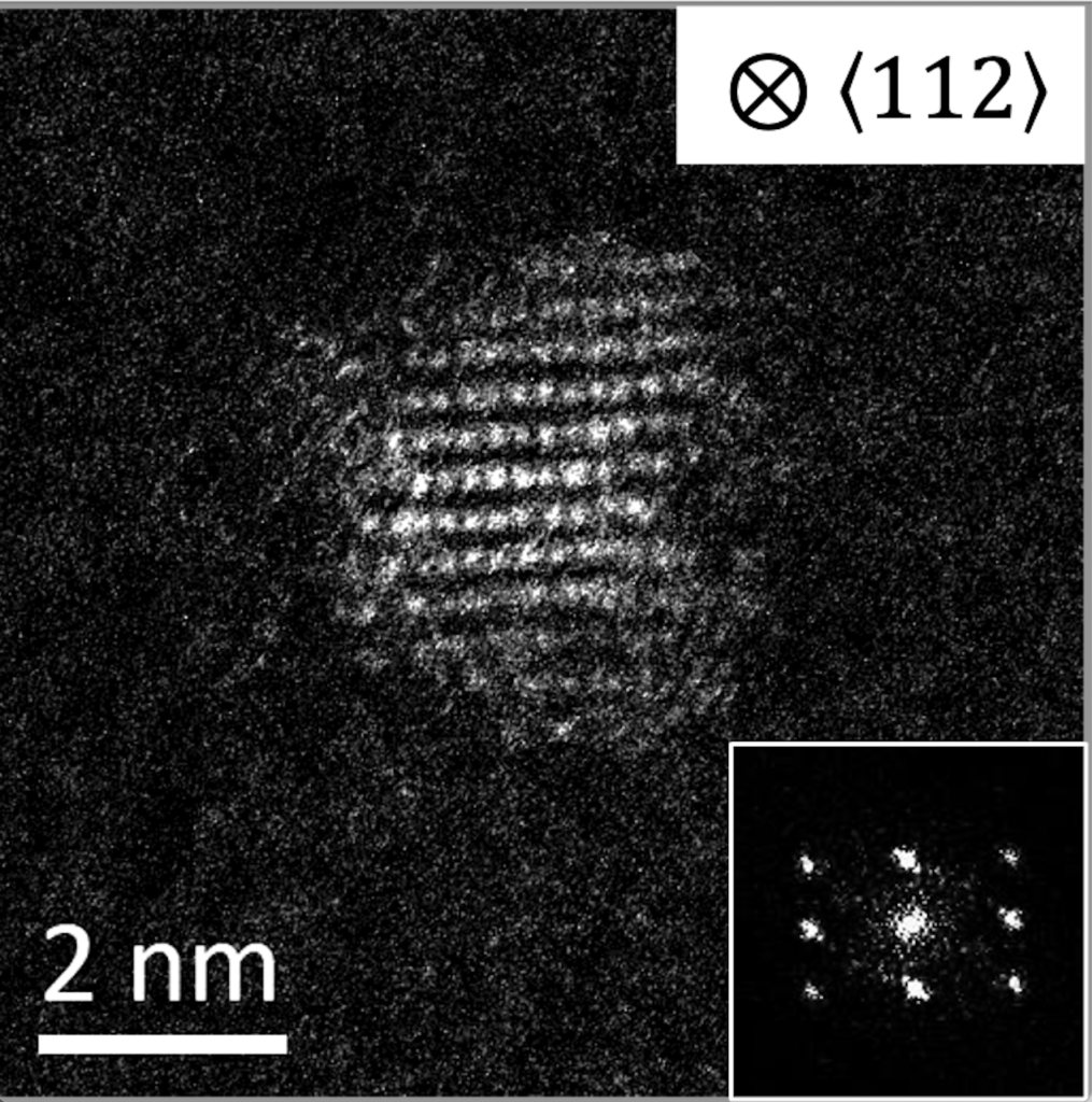
Imaged under an electron microscope, these quantum dots were produced in the Hecht Lab using de novo proteins. Each quantum dot is 2 nanometers in diameter, an important factor since particle size determines the color they glow, or fluoresce, in.
Researchers found that ConK enabled the survival of E. coli in otherwise toxic concentrations of copper, suggesting it might be useful for metal binding and sequestration. The quantum dots used in this research are made out of cadmium sulfide. Cadmium is a metal, so researchers considered ConK as they sought new ways to synthesize quantum dots.
Their hunch paid off. ConK breaks down cysteine, one of the 20 amino acids, into several products, including hydrogen sulfide. That acts as the active sulfur source that will then go on to react with the metal cadmium. The result is CdS quantum dots.
“To make a cadmium sulfide quantum dot, you need the cadmium source and the sulfur source to react in solution,” said Spangler. “What the protein does is make the sulfur source slowly over time. So, we add the cadmium initially but the protein generates the sulfur, which then reacts to make distinct sizes of quantum dots.”
The growth of the quantum dot particle is very slow, making it easy to interrupt the process at any point to control for size. With quantum dots, particle size determines the color the dots fluoresce in: the smaller particles fluoresce blue, for example; the larger ones, red.
“They can be useful for anything that you want a fluorescent tag for,” said Spangler, who now runs her own lab at Virginia Commonwealth University. “In one of my previous papers, we attached an antibody to the quantum dots so that they would mark specific cancer cells. Once they are marked, you can look under a microscope and only the cancer cells light up.”
For the full paper in PNAS, click here. To view a video on this research prepared by Princeton Innovation, click here.
“A de novo protein catalyzes the synthesis of semiconductor quantum dots,” was authored by Leah Spangler, Yueyu Yao, Guangming Cheng, Nan Yao, Sarangan Chari, Gregory Scholes, and Michael Hecht. The research was supported by the National Science Foundation MRSEC program (DMR-2011750). Additional funding was providing by the Princeton University Writing Center, and the Canadian Institute for Advanced Research. The research was also supported by NSF grant MCB-1947720 to MH.