Rabitz, Jonikas Awarded $3.4M Moore Foundation Grant
In a funding venture that could be transformational for imaging single molecules within a cell, the Gordon and Betty Moore Foundation has awarded a $3.4M grant to a collaboration between Princeton’s Departments of Chemistry and Molecular Biology.
The four-year grant supports the development of a new microscope that will enable researchers unprecedented access to the chemical composition and distribution of small molecules within cells, at nanometer resolution.
At present, there is no technology capable of achieving high-resolution imaging and chemical species identification within a cell.
The co-principal investigators on the team include chemistry’s Herschel Rabitz, the Charles Phelps Smyth ’16 *17 Professor of Chemistry; and Associate Professor of Molecular Biology Martin Jonikas, also an investigator with the Howard Hughes Medical Institute.
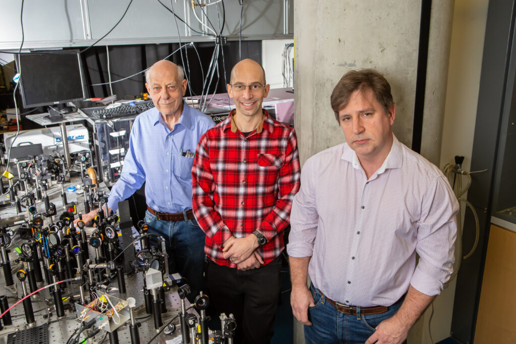
Grantees (l to r): Herschel Rabitz of Princeton Chemistry, Martin Jonikas of Princeton Molecular Biology, with Alexei Goun of Princeton Chemistry.
Their microscope will draw on infrared vibrational spectroscopy and quantum dynamics to characterize the chemical nature of each molecule. Knowing where the small molecules are within the cell and what they’re doing will advance basic scientific understanding of cellular biology that, in turn, will push the frontier of therapeutics.
Researchers will build the instrument at Princeton Chemistry using a sophisticated laser system and components being shipped from vendors all over the world. Once assembled, it will operate at a “sweet spot” of a few nanometers, a level of resolution that will allow for chemical identification.
“The email about the grant came to me late at night with a message from the program manager at the foundation. It was very exciting,” said Rabitz. “It is refreshing that the foundation is not intimidated by this research because it is going to be a challenge. They’re attracted to things that are transformative. And things that are transformative are invariably difficult to achieve.
“The goal of this project rides on quantum,” Rabitz added, “but it will be an integrated instrument that uses a sharp tip to achieve images smaller than the wavelength of the light—a technique called tip-enhancement spectroscopy—and employs quantum effects to draw out unique signatures from each chemical species. That is, we will be able to see where the molecules are located and what they are.”
Added Jonikas: “We’re going to combine existing technologies in ways that haven’t been tried before. There are unknowns about exactly how well our planned approaches will work, and there’s some risk of unexpected challenges.
“But I think there is also a huge space of opportunity to work within. Whatever challenges we encounter, we should be able to overcome.”
The overall concept of the instrument is credited to Alexei Goun, a former research specialist with the Rabitz Group who came up with the idea after listening to Jonikas’ research flash talk at the 2018 Princeton Catalysis Initiative Symposium.
The research team also includes postdoc Katerina Kanevche, likely the first person in the world to take a 3D image of a single cell using tip-enhanced spectroscopy (TES). The team is rounded out by second-year graduate student Amr Sobeh. Both Kanevche and Sobeth are jointly advised by Rabitz and Jonikas.
The team has already received funding from the Eric and Wendy Schmidt Transformative Technology Fund and the Howard Hughes Medical Institute.
Approaching the Challenge
With their new technology, researchers seek to probe the biochemistry inside a cell—which contains thousands of small molecules—both under normal conditions and in disease states.
The first step in development includes gathering hundreds of components, a process with many challenges. For one thing, the components were not developed for integration into one instrument. Many pieces had to be redesigned after months of discussion among vendors from multiple countries. Assembly will likely generate unforeseen complications.
But researchers envision a “transformative” instrument that can spectroscopically identify and distinguish one molecule from another, and then determine its chemical makeup.
“We need to determine the chemical specificity, and this is what’s unique about this instrument,” said Hersch. “It will be able to tell us exactly what biochemicals we’re looking at.”
Once the instrument is assembled, the beta test for its capabilities will be Jonikas’ own research, which focuses on CO2 assimilation through photosynthesis in a certain species of algae. Researchers believe algae enhance this assimilation by pumping CO2 to a high concentration in a compartment within the cell – a spatial distribution of CO2.
Currently, it’s impossible to see that distribution. This is the point at which Goun’s idea took shape four years ago: if researchers could assemble an imaging device that could look inside a cell, they could gain insights into how this assimilation works.
Similar challenges exist for the characterization of hundreds of cellular metabolites. Colloquially, the term “metabolite” refers to any small molecule within a cell.
“That’s where this new instrument enters the picture,” said Jonikas. “It will really be the first general technology to look at any metabolite. And currently, for CO2, we know of no other viable candidate technology.
“That’s why we’re so excited about this instrument.”
This research is funded by the Gordon and Betty More Foundation through Grant GBMF 12215. The Gordon and Betty Moore Foundation fosters path-breaking scientific discovery, environmental conservation, patient care improvements, and preservation of the special character of the Bay Area.