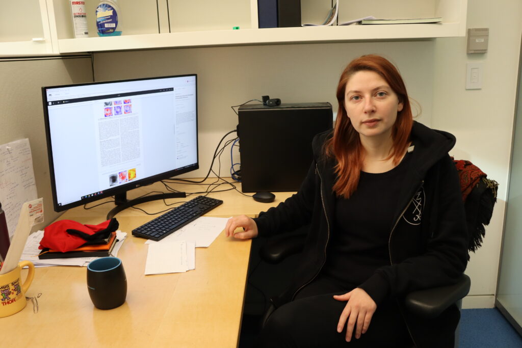
Postdoc Kanevche heads up groundbreaking microscope project to ID chemical species

From the 30K-foot level, Katerina Kanevche moved effortlessly from graduate student to postdoc to leader on a project whose goal is a novel imaging and molecular identification instrument that could transform the field of microscopy.
In reality, she painstakingly acquired experience in a technique called tip-enhanced spectroscopy (TES), gaining skills that landed her among an elite group of practitioners around the world.
And while the opportunity to lead the project at Princeton Chemistry fell into her lap when a former researcher departed the program, her appointment has all the earmarks of an outcome that was meant to be.
TES combines optical, laser-based microscopy and spectroscopy with scanning probe technology for a new level of imaging power without the limitations of diffraction. Kanevche honed her expertise in this area in Germany at the Freie Universität Berlin where she earned her Ph.D. in physics in March 2022. There, she employed this technique to record infrared images revealing a cell’s subcellular structures and constructed the world’s first 3D image of the local protein content within the cell’s organelles.
The world’s first 3D image of the local protein content within a cell’s organelles, which Kanevche obtained through multiple cross-sections using her skills in tip-enhanced spectroscopy.
Today, Kanevche is a postdoc in the Rabitz Lab, recruited to Princeton to round out the team of researchers who plan to build a first-of-its-kind microscope that will image small molecules within a cell and simultaneously determine their chemical composition.
“Several things just fit together at the perfect time,” said Kanevche from her office at Frick Lab. “When I started out with my Ph.D. project, I was eager to learn new things and was excited to be involved in such challenging and cutting-edge research. As I dived deeper and realized the enormous potential of this research in biophysics, my passion and drive only grew.
“After all, to a large extent, I have performed proof-of-concept experiments. But high-impact applications are still to come, especially from combining imaging with chemical identification of the species being observed. I am so very lucky and excited to both witness and be a part of these developments.”
Kanevche is working under Herschel Rabitz, the Charles Phelps Smyth ’16 *17 Professor of Chemistry, and Associate Professor of Molecular Biology Martin Jonikas, both of whom have been named co-P.I.s on several landmark grants funding the project. The team also includes graduate student Amr Sobeh, who is jointly advised by Rabitz and Jonikas.
Researchers anticipate the new instrument will be applicable to all sorts of biophysical problems, from investigating cancer cells with exceptional detail to elucidating biophysical or biochemical mechanisms.
Rabitz said Kanevche’s “commanding presence,” evident during her first interview with P.I.s last spring, made her a natural choice to lead the project.
“Katerina has eagerly taken the reins of overseeing the search for scientists to form an integrated microscope construction team,” said Rabitz. “Her approach to constructing the microscope comes from the perspective of enabling a deeper understanding of biology at the fundamental molecular level within a single cell. Her leadership will surely play a key role in guiding the team to solve the myriad of technical challenges ahead.”
A New Kind of Microscope
Last fall, the Rabitz-Jonikas team received a multimillion-dollar grant from the Gordon and Betty Moore Foundation to develop a microscope that will enable researchers unprecedented access to the chemical composition and distribution of small molecules within cells. There are techniques that image small molecules and techniques that determine the chemical makeup of small molecules; but nothing, to date, that combines them at a spatial resolution below 20 nanometers.
Their project looks to achieve this goal.
Researchers will use a kind of TES called scattering-type scanning near-field optical microscopy (sSNOM) to overcome diffraction limitations. The microscope includes a sophisticated laser system working within a cell at 4-degrees Kelvin to build an instrument that uses high resolution infrared vibrational spectroscopy combined with high sensitivity quantum dynamics signatures to identify the molecules being viewed in the cell.
Assembly itself is likely to be a challenge, with a myriad of possible pitfalls to overcome and opportunities for innovations along the way. Kanevche said these factors were among the reasons she was intrigued by the project.
Two years ago, along with colleagues at Free University, Kanevche was first author on a paper in Communications Biology that detailed the world’s first 3D image of the subcellular infrared absorption of proteins, a composite of cross-sections of the protein distribution inside a green algae cell. The resolution for this work was about one-hundred-fold better than with conventional infrared microscopes.
The paper announced her as a player in this growing field, with s-SNOM as her specialty.
“s-SNOM is one type of tip-enhanced technique where laser radiation—in my case infrared radiation—is focused on an atomic force microscopy (AFM) tip,” she said. “This approach allows us to overcome the diffraction limit that normally sets a fundamental limitation to the spatial resolution.
“The highlight of this research is that we had chemical images that reveal, for example, the protein distribution on a given cell area. The recorded images reported in the paper were with spatial resolution of 20 nanometers. This is not possible to do with any conventional infrared microscope, anywhere. It was very, very exciting.”
Kanevche will bring these skills to bear as she leads the Princeton-based project. The plan is to take the chemical specificity offered by optical high resolution spectroscopy along with high sensitivity molecular quantum dynamics signatures and combine them with atomic force microscopy. (Atomic force microscopy involves a very small, sharp tip that can scan a cell’s surface through interactions between the atoms of the tip and the atoms on the surface of the cell.)
By combining these two techniques, researchers can get chemical specificity with the resolution of atomic force microscopy. Depending on the laser source used, researchers can acquire images or chemical specificity with spatial resolution comparable to the size of the AFM tip.
“This research matches perfectly with my interests and profile as a scientist, and at the same time introduces several layers of novelty and complexity that make it very challenging and exciting,” Kanevche concluded. “I believe I can guide the progress and development of this project and invest my entire experience toward reaching our goals.”
